Single Transducer Probe: Physics, Types, and Clinical Applications
Background
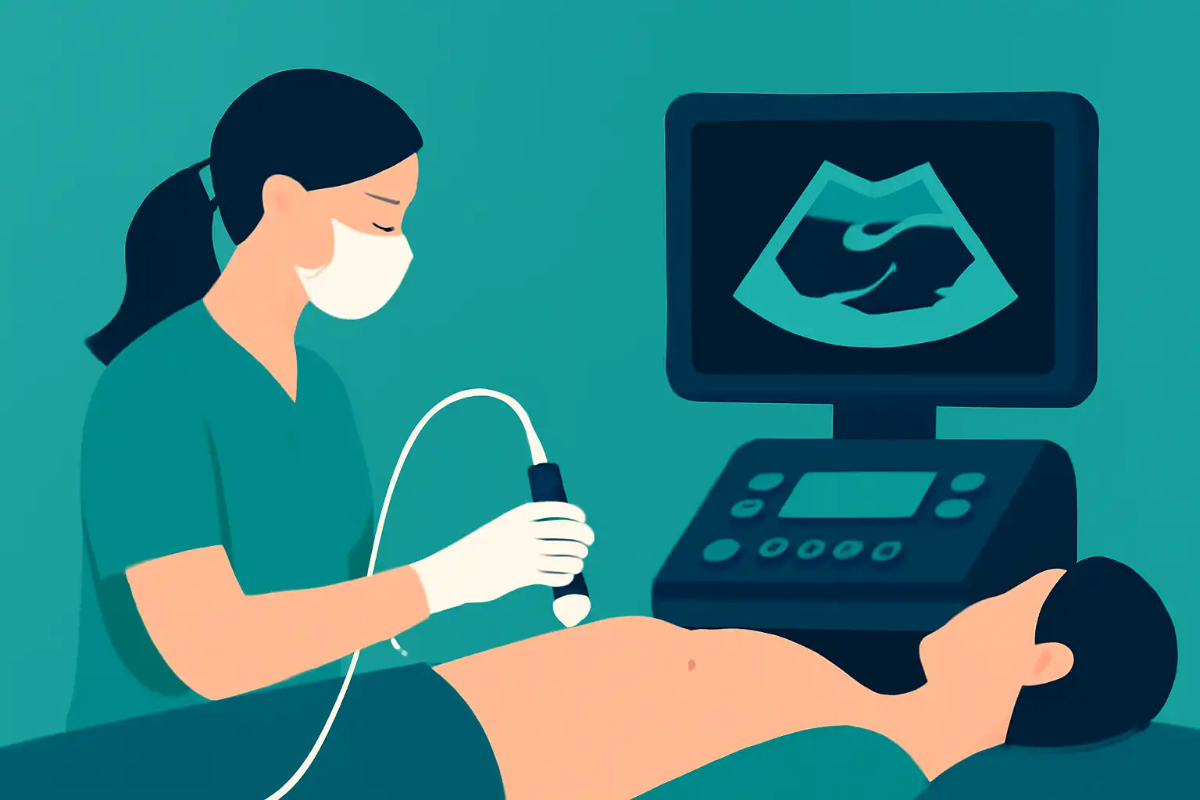
A single transducer probe is the fundamental handheld device used in diagnostic ultrasound. It contains one array of piezoelectric elements that converts electrical energy into acoustic energy (and vice versa) to generate and receive sound waves. Its design dictates the shape of the ultrasound beam, the field of view, and the optimal depth of imaging, making it a critical determinant of image quality and clinical utility.
Core Physics & Components:
- Piezoelectric Elements: Crystals or ceramic materials that vibrate when an alternating current is applied, producing sound waves at a specific frequency. The received echoes cause them to generate an electrical signal.
- Backing Material: Dampens the crystal’s vibration to improve axial resolution (shorter pulse length).
- Matching Layer: Reduces the acoustic impedance mismatch between the crystal and the skin, allowing more sound energy to enter the body.
- Acoustic Lens: Focuses the sound beam in the elevation plane (slice thickness).
The performance of any single transducer is governed by trade-offs, primarily between frequency and depth.
- Higher Frequency (e.g., 7-15 MHz): Provides superior resolution but has greater attenuation, limiting depth of penetration. Ideal for superficial structures (thyroid, breast, MSK).
- Lower Frequency (e.g., 1-5 MHz): Provides less resolution but better penetration for deeper structures (abdomen, obstetrics).
Common Single Transducer Probe Types:
| Probe Type (Array) | Beam Shape & Field of View | Typical Applications |
|---|---|---|
| Linear Array | Rectangular / Parallel | MSK, Vascular, Small Parts, Thyroid |
| Curved (Convex) Array | Trapezoidal / Sector with wide near field | Abdominal, Pelvic, Obstetric |
| Phased Array | Narrow sector with small footprint | Cardiac, Transcranial (through acoustic windows) |
What’s New
While the basic physics of piezoelectric transduction is well-established, recent advances focus on materials science, integration, and processing to enhance the capabilities of single probes.
- Advanced Materials & Fabrication:
- Single Crystal Piezoelectrics: Increasingly used in high-end probes, these offer higher electromechanical coupling efficiency and bandwidth than traditional PZT ceramics. This results in improved sensitivity and image resolution.
- MEMS Technology: Capacitive Micromachined Ultrasound Transducers (CMUTs) are an emerging class. They offer wide bandwidth, ease of integration with electronics, and potential for miniaturization. Their clinical adoption is growing.
- Wideband & Harmonic Imaging: Modern probes are designed as wideband transducers, capable of operating over a broad frequency range. This is essential for:
- Tissue Harmonic Imaging (THI): Now a standard feature on virtually all probes. It uses non-linear propagation effects to image at twice the fundamental frequency, significantly improving contrast resolution and reducing artifact.
- Integration and Ergonomics:
- Beamformer-on-Probe: Processing is increasingly moved into the probe handle, allowing for better signal-to-noise ratio and cable efficiency.
- Multi-Parametric & Fusion Probes: While still single transducer devices, new probes are designed with integrated sensors for elastography (shear wave) or are optimized for fusion with CT/MRI datasets.
Clinical & Practice Impact
The selection and understanding of a single transducer probe is a daily clinical decision that directly impacts diagnostic accuracy, workflow, and patient safety.
- Probe Selection is a Primary Skill: Choosing the wrong probe type or frequency is a common cause of suboptimal imaging. A clinician must match the probe to the anatomical region and depth of the target structure.
- Optimizing Image Quality: Understanding the physics allows for informed adjustments. For example, knowing that a higher frequency improves resolution can guide a user to change probes or increase the frequency setting when examining a superficial, poorly defined lesion.
- Artifact Recognition: Many common artifacts are probe-related. Side lobe artifacts and slice thickness artifacts are directly related to the beam formation of the single transducer array. Recognizing these as probe-based phenomena prevents misdiagnosis.
- Impact on Specific Exams:
- Cardiac (Phased Array): Its small footprint and ability to steer the beam through acoustic windows (intercostal spaces) are non-negotiable for standard echocardiography.
- Musculoskeletal (Linear Array): High-frequency linear probes are essential for detailed resolution of tendons, nerves, and small joints.
- Abdominal (Curved Array): The wide field of view is necessary to survey large organs and track pathology in relation to surrounding anatomy.
Probe-Driven Diagnostic Trade-offs:
| Clinical Scenario | Ideal Probe Choice | Rationale & Trade-off |
|---|---|---|
| Superficial soft tissue lump | High-frequency Linear (>10 MHz) | Gain: Maximum resolution to characterize margins and internal texture. Loss: Limited depth; cannot assess deep extension. |
| Deep right upper quadrant pain | Low-mid frequency Curved Array (2-5 MHz) | Gain: Penetration to visualize liver, gallbladder, kidney. Loss: Lower spatial resolution compared to a linear probe. |
| Assessing fetal heart in 2nd trimester | Low-frequency Curved Array OR Phased Array | Gain: Adequate penetration through maternal abdomen. The choice may depend on maternal habitus and fetal position. |
Bottom Line
The single transducer probe remains the cornerstone of diagnostic ultrasound. Its design—a direct consequence of fundamental ultrasound physics—dictates clinical capability.
- Fundamentals are Key: Proficiency requires understanding the direct relationship between frequency, resolution, and penetration and how this is embodied in the three core probe geometries (Linear, Curved, Phased).
- Technology is Evolving: Advances in materials (single crystal, CMUTs) and processing (wideband, harmonic imaging) are steadily improving image quality, but do not change the core physical trade-offs clinicians must navigate.
- Clinical Impact is Direct: Appropriate probe selection is the first and most critical step in any ultrasound examination. A mismatch leads to poor image quality, misinterpretation, and increased scanning time.
- For the Exams (ARDMS SPI, FRCR Part 1): A robust understanding of transducer construction, beam properties (focusing, steering), and the physical basis for choosing one probe type over another is essential. This knowledge underpins both the physics questions and the image interpretation required in clinical practice.
Discussion & Comments