Cellular Adaptation in Pathology: Board Review of Atrophy, Hypertrophy, Hyperplasia, Metaplasia, and Dysplasia
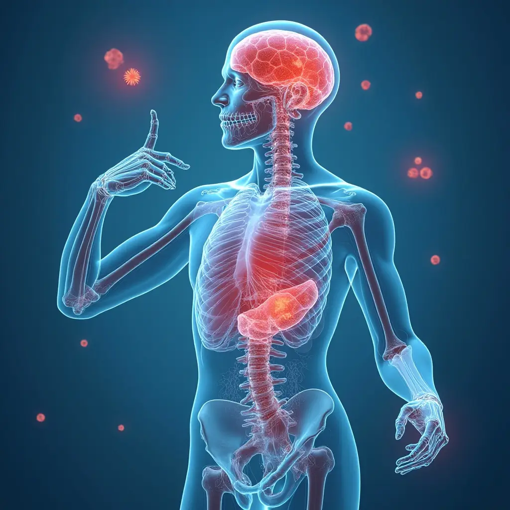
CME & Learning (Board Review) Article: Cellular Adaptation
Overview
Cellular adaptation is a reversible, structural and functional response of cells to changes in their environment. These adaptations allow cells to survive in altered physiological states or to withstand pathological stimuli. The type of adaptation depends on the nature, severity, and duration of the stimulus, as well as the cell’s capacity to adapt. Key adaptations include atrophy, hypertrophy, hyperplasia, metaplasia, and dysplasia. Understanding these concepts is fundamental for pathology and is heavily tested on USMLE Step 1 and AMC MCQ exams.
High-Yield Key Points
- Atrophy: A decrease in cell size and function. Results from decreased workload, loss of innervation, diminished blood supply, inadequate nutrition, loss of endocrine stimulation, or pressure. Due to increased autophagy (degradation of cellular components) and ubiquitin-proteasome pathway activity.
- Hypertrophy: An increase in cell size. Common in permanent cells (e.g., cardiac myocytes, neurons) that cannot divide. Driven by growth factors and increased protein synthesis. Examples: left ventricular hypertrophy in hypertension, uterine enlargement in pregnancy.
- Hyperplasia: An increase in cell number. Occurs in labile and stable cells that retain the ability to divide. Caused by hormonal or growth factor stimulation. Example: endometrial hyperplasia due to estrogen, lymph node hyperplasia during infection.
- Metaplasia: A reversible change of one differentiated cell type to another. A direct stem cell reprogramming adaptation to chronic stress, often involving exposure to irritants (e.g., smoke, acid reflux). Example: Barrett’s esophagus (squamous to columnar epithelium). Important: Metaplasia can progress to dysplasia and carcinoma.
- Dysplasia: Not a true adaptation but a disordered proliferation, often considered a pre-neoplastic lesion. Characterized by loss of uniformity and architectural orientation of cells. It is reversible if the stimulus is removed, except when it becomes high-grade/in situ carcinoma. Example: cervical intraepithelial neoplasia (CIN).
Mnemonics
- Mnemonic for the Four Main Reversible Adaptations: “HHAH”
- Hypertrophy (Bigger cells)
- Hyperplasia (More cells)
- Atrophy (Smaller cells)
- Hypoplasia/Metaplasia (Underdeveloped/Changed cells)
Comparison Tables
| Feature | Hypertrophy | Hyperplasia |
|---|---|---|
| Primary Change | ↑ Cell size | ↑ Cell number |
| Cell Type | Permanent cells (myocytes, neurons) | Labile/Stable cells (epithelial, fibroblasts) |
| Mechanism | ↑ Protein synthesis, organelle size | ↑ Cell proliferation via mitosis |
| Key Stimulus | Increased functional demand, hormonal | Hormonal, growth factors, chronic injury |
| Example | Cardiac myocyte in hypertension | Hepatocytes after partial hepatectomy |
| Feature | Metaplasia | Dysplasia |
|---|---|---|
| Definition | Replacement of one differentiated cell type by another | Disordered, atypical cell growth |
| Nature | Adaptive, reversible | Pre-neoplastic, potentially reversible |
| Cause | Chronic irritation/inflammation | Often follows persistent metaplasia/irritation |
| Cell Appearance | Mature, differentiated cells (but wrong type) | Immature, pleomorphic, hyperchromatic cells |
| Malignant Potential | Low, but a precursor step | High if high-grade; can progress to carcinoma |
| Example | Squamous metaplasia in bronchi of smokers | Cervical dysplasia (CIN) |
Board-Style Questions
Question 1 A 55-year-old male with a long-standing history of uncontrolled hypertension presents with a new diagnosis of congestive heart failure. An echocardiogram reveals a significantly thickened left ventricular wall. Which of the following cellular adaptations is the primary mechanism for the increased ventricular wall thickness? A) Hyperplasia of cardiac myocytes B) Hypertrophy of cardiac myocytes C) Metaplasia of the endocardium D) Fibrosis of the myocardium
Answer: B) Hypertrophy of cardiac myocytes. Explanation: Cardiac myocytes are permanent cells and cannot undergo hyperplasia. In response to increased workload (from hypertension), they adapt by increasing their size (hypertrophy), which involves increased protein synthesis and organelle size. This leads to ventricular wall thickening. Fibrosis (D) is a form of scarring/injury, not a primary adaptive response to increased pressure. Hyperplasia (A) is incorrect for myocytes. Metaplasia (C) is not relevant here.
Question 2 A gastroenterologist performs an endoscopy on a patient with chronic, severe gastroesophageal reflux disease (GERD). A biopsy of the lower esophagus reveals intestinal-type goblet cells in an area normally lined by stratified squamous epithelium. Which of the following terms best describes this finding? A) Dysplasia B) Hyperplasia C) Metaplasia D) Atrophy
Answer: C) Metaplasia. Explanation: This describes Barrett’s esophagus, a classic example of metaplasia. The chronic acid injury causes the normal squamous epithelium of the esophagus to be replaced by a more acid-resistant columnar epithelium with goblet cells (intestinal metaplasia). It is a reversible change (if stimulus removed) but is a known risk factor for adenocarcinoma. It is not dysplasia (A), which implies disordered atypia. Hyperplasia (B) is an increase in cell number, not a change in type. Atrophy (D) is a decrease in cell size.
Question 3 A patient with chronic obstructive pulmonary disease (COPD) has a biopsy of the bronchial epithelium. The pathologist notes a replacement of the normal pseudostratified ciliated columnar epithelium by stratified squamous epithelium. This change is: A) An adaptive response that increases the epithelial barrier to infection B) A direct precursor to small cell carcinoma C) A reversible change caused by chronic cigarette smoke exposure D) A form of neoplastic transformation
Answer: C) A reversible change caused by chronic cigarette smoke exposure. Explanation: This is squamous metaplasia of the bronchial epithelium, a common response to chronic irritation from cigarette smoke. It is a reversible adaptive change. However, the new squamous cells are more susceptible to dysplasia and eventual squamous cell carcinoma, not small cell carcinoma (B). While it is an adaptation, it is not primarily to enhance barrier function (A) as cilia are lost, impairing clearance. It is not neoplastic (D) at this stage.
Question 4 Which of the following cellular adaptations is least likely to be reversible? A) Hepatocyte hyperplasia following partial hepatectomy B) Endometrial hyperplasia due to unopposed estrogen C) High-grade cervical dysplasia D) Uterine smooth muscle hypertrophy during pregnancy
Answer: C) High-grade cervical dysplasia. Explanation: While dysplasia is often reversible when the inciting stimulus is removed, high-grade dysplasia (CIN III) is considered synonymous with carcinoma-in-situ and represents a direct precursor to invasive cancer. It is often effectively treated by excision (e.g., LEEP procedure) but is fundamentally a neoplastic, rather than a truly reversible adaptive, process. The other choices (A, B, D) are classic, reversible physiological or pathological adaptations.
Summary
- Cellular adaptations are reversible responses to stress, allowing cell survival.
- Key types: Atrophy (↓ size), Hypertrophy (↑ size), Hyperplasia (↑ number), Metaplasia (cell type change).
- Cardiac myocytes undergo hypertrophy, not hyperplasia.
- Metaplasia (e.g., Barrett’s, squamous bronchial) is a critical precursor to dysplasia and carcinoma in chronic injury settings.
- Dysplasia is a disordered growth pattern with pre-neoplastic potential; high-grade dysplasia borders on carcinoma.
- Understanding the difference between these adaptations and their clinical contexts is essential for pathology questions on board exams.
Discussion & Comments